|
|
Magneto-optical spectroscopy laboratory |
|
Budapest University of Technology and Economics
Faculty of Natural Sciences
Institute of Physics
Motivation of the research
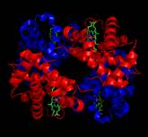
Optical spectroscopy on complex magnetic materials gains increasing research potential due to the fundamental and detailed information recently obtained about the character of the magnetic ordering in many systems. Beyond the bulk magnetization, the method often allows the determination of the magnetization for the different components or sublattices. Futhermore, basic physical parameters such as the exchange and spin-orbit couplings can be also deduced from the magneto-optical (MO) spectra. The broadband MO spectrometer developed in our laboratory offers a solid experimental background to study novel and technologically important magnetic materials on a broad length-scale, down to low temperatures and in high magnetic fields. Beyond the research of inorganic magnets, we also investigate biological systems by polarized optical and magneto-optical spectroscopy. Our main topics in this field are the secondary structure analysis of proteins by circular dichrosim spectroscopy and the magneto-optical diagnosis of malaria.
Experimental facilities
Most of the MO measurement systems used in our laboratory have been – partly or fully – developed by our group. Based on these facilities we can detect reflectivity, transmission, and magneto-optical parameters – including magneto-optical Kerr effect (MOKE), Faraday effect, magnetic and natural circular dichroism – over the spectral range of 0.1-7eV (170-12000nm in wavelength). The high-sensitivity magneto-optical and natural circular dichroism experiments are developed with the application of a polarization modulation technique using fast photodetectors, photoelastic modulators and lock-in amplifiers.
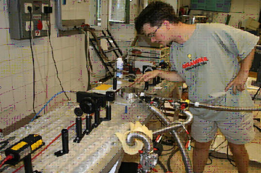
· Low-temperature MO measurement unit
|
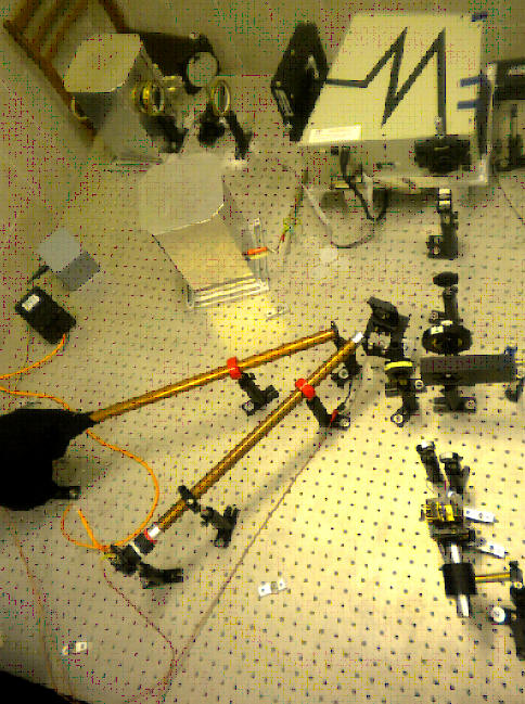
This facility is composed of infrared and visible laser diodes, polarizing optical elements and a photoeleastic modulator. Experiments can be performed in the temperature range of T=2-300K using a continuous-flow liquid helium optical cryostat. The precision of the detection for the MO parameters is typically ≤0.001°. Magnetic field of B=0.4T in maximum is applied by NdFeB permanent magnets.
|
|
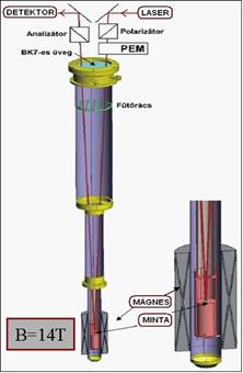
· High-field MO measurement system
Concerning the building blocks this system is similar to the low-temperature MO unit. The substantial difference is that it can be integrated with a high-field, B=14 T, supercondacting magnet. Using a home-designed optical insert the sample is also loaded into the liquid helium cryostat and placed to center position within the superconducting magnet. The whole light path is realized using optical elements. The temperature of the samples can be varied over the range of T=2-300 K.
 |
 |
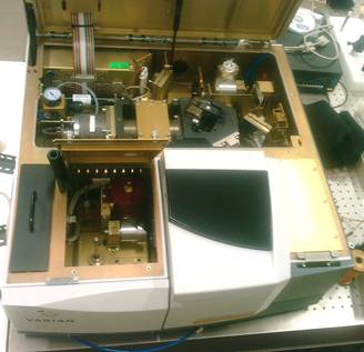
· Broadband MO spectrometer
The MO spectrometer developed in our laboratory with a uniquely broad spectral range (0.1-7 eV) is built on the basis of two optical spectrometer. For lower photon energies (0.1-0.9 eV) we adapted the polarization modulation technique for our custom-designed Varian-670 two-channel Fourier transform infrared spectrometer. For higher-energy (0.7-7 eV) studies a Cornerstone ¼ monochromator is applied with three interchangeable holographic gratings. In order to cover such a broad spectral region we use various types of detectors such as MCT, InSb, InGaAs, Si photodiodes and a photomultiplier. A resolution of ~0.001° for the magneto-optical parameters is achieved over the whole range of photon energy specified above. This MO spectrometer also allows low-temperature experiments in magnetic field of B=0.4 T in maximum.
 |
 |
Main research topics
·
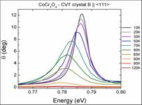
Complex form of
magnetic ordering studied by MO spectroscopy
|
|
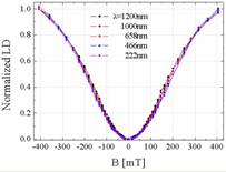
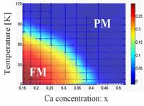
MO spectroscopy can often reveal much detailed information about the magnetic structure and the character of the electrons responsible for the magnetism as compared with bulk magnetization experiments, especially in case of magnets with various magnetic components or sublattices. For some materials case we also investigated the magnetic phase diagram using composition, temperature and pressure as control parameters.
|
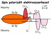
·
Mo spectroscopy
on itinerant magnets
|
|
Determination of spin-polarized band structure in itinerant ferromagnets is challenging both from experimental and theoretical side and would help the synthesis of completely spin-polarized conductors. For several metals we could deduce the accurate spin-polarized electronic structure from the simultaneous measurements of absorption and magneto-optical spectra which give complementary information about the nature of the electronic states. |